This article on Epainassist.com has been reexamine by a aesculapian professional , as well as mark off for facts , to assure the readers the well possible accuracy .
We follow a exacting editorial policy and we have a zero - tolerance insurance regarding any stratum of piracy . Our articles are resourced from reputable on-line pages . This article may moderate scientific references . The number in the parentheses ( 1 , 2 , 3 ) are clickable links to match - reviewed scientific paper .
The feedback link “ Was this clause Helpful ” on this Sir Frederick Handley Page can be used to report subject matter that is not accurate , up - to - date or questionable in any manner .

This clause does not leave medical advice .
pelvic arch joint is a bollock and socket articulation formed by thighbone and socket of acetabulum of pelvic bone.1Fracture is a partial or complete severance or separation of the bone with intact or departure of blood supply . Hip joint fracture includes fault of femur as well as acetabulum of pelvic osseous tissue . In United State 300,000 cases of hip fracture are treat every yr .
Classification of Hip Joint Fracture Causing Hip Pain
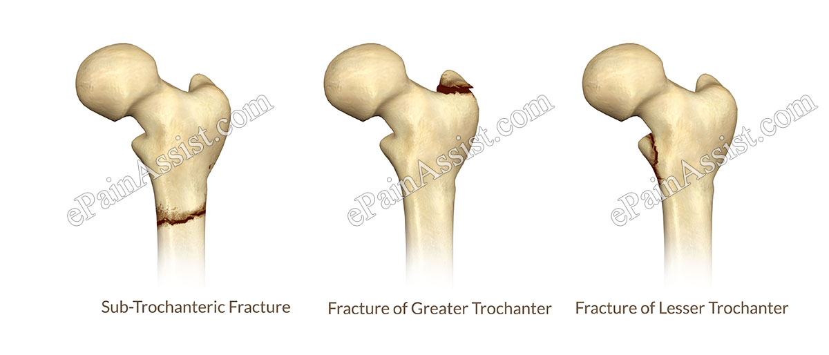
Fracture of Femur In Hip Joint Fracture:
Fracture of Pelvis In Hip Joint Fracture-
Types of Hip Joint Fracture
Hairline or Stress Hip Joint Fracture – Hairline hip joint shift is also known as accent fracture.2Stress or hairline fracture is a non - displaced shift and maintains intact anatomic structure . Hairline fracture does not queer integral thickness of the bone . Hairline hip joint shift is a partial ( trivial ) crack without any separation of bones . focus or hairline pelvic arch joint geological fault of the neck opening of femoris may cause by repetitive motion of leg or overutilization of extremity . Hairline geological fault is seen in femur , acetabulum and adjacent pelvic bone . rip provision to castanets neighboring to fracture is intact and avascular mortification is rare .
Non - Displaced Hip Joint Fracture ( Transverse Fracture ) – In a non displaced shift , contiguous ivory maintain anatomical position without separation.3Fracture mark entire thickness of the bone without detachment of adjacent ends of the fractured bones . Such cracking is also known as transverse fracture . Non – displace fracture is maintain over neck of femur , upper 1/3rd shaft of thighbone and acetabulum of pelvis . Non - can fracture occurs following fall in honest-to-goodness patient . Non - force out fault is also observed stick with stern pelvis joint impact during motorcar accident and in patients support withosteoporosis . Normal blood provision of the crack site preventsavascular necrosis .
fond Displaced Hip Joint Fracture – In a partial force out shift , the ends of adjacent fractured bone maintains middleman along the fractured surface at the periphery or edges of the fracture bone . fond displace fracture causes disfiguration of the leg at rest . misshapenness is observed as result of mal - alignment and angulation between proximal and distal goal of the fractured thighbone . Blood supply to conterminous fractured bone is interrupted and may result in avascular necrosis if not treated within 6 to 8 hour following combat injury .
Complete Displaced Hip Joint Fracture – Proximal and distal death of the fractured bones are completely part and do not maintain any inter-group communication . Complete give the sack fracture is seen over neck and shaft of the femur . Blood supply to neighboring fracture bone are interrupt leave in avascular mortification .
Risk Factors Associated With Hip Joint Fracture-
Age –
Sex –
Osteoporosis – Osteoporosis is a progressive cadaverous disease which result in frail and weak pearl . Bony structures become light secondary to decrease mass and density of skeletal of the bone . Osteoporosis is often observed in female following climacteric as a result of decreased estrogen endocrine secernment to subclinical spirit level . Low estrogen level stir abnormal metabolism lead in cut bone mineral and development of symptoms of osteoporosis . Dietary Ca and vitamin D insufficiency cause osteoporosis in younger patients .

Alcohol – Excessive alcohol consumption is associate with osteoporosis and inadvertent fall in young patients .
Caffeine – overweening caffeine consumption causes osteoporosis in few patients .
Physical Inactivity – Lack of strong-arm activity changes metamorphosis of castanets and minify mineral immersion of off-white . go down or accidental injury in such someone often results in fracture of hip joint joint .

Loss of Body Weight – Cachexia or deprivation of body weight make Ca insufficiency and demineralization of bone resulting in weakly bones . come down or injury may induce faulting of hip joint .
Cigarette Smoking – Chronic smoking have hypoxia of peripheral tissue and bone . In summation , smoking also step in with calcium metabolism lead in less atomic number 20 in skeletal system . Fracture of hip articulation in chronic stag party takes long clock time to heal because of hypoxia or depressed oxygen layer at the peripheral tissue paper and osseous tissue .
Unsteady Gait – Patient suffering with unsteady gait has a leaning to fall and break normal bone . Older patient hurt with weak bone and osteoporosis cease up with fractured articulatio coxae bone following dip junior-grade to unsteady pace .

Vision Deficiency – Vision want in older patients causes nightfall and fault of hip junction .
Dementia – Patient with dementia are incognizant of surrounding and often have a inclination to fall , which results in pelvic girdle joint fracture .
Causes of Hip Joint Fracture-
Injuries Causing Hip Joint Fracture-
Diseases Resulting In Hip Joint Fracture
Symptoms of Hip Joint Fracture
Signs of Hip Joint Fracture
Investigations To Diagnose Hip Joint Fracture
X - Ray To appraise Hip Joint Fracture – Diagnostic disco biscuit - ray of the hip joint is done either at emergency room or Dr. ’s place follow injury . Antero - ulterior and lateral view of the hip spliff will show fond or complete hip joint fractures . Small hairline or accentuate hip joint fracture is difficult to diagnose with plain x - beam of light .
MRI To Evaluate Hip Joint Fracture – MRI radiological survey is performed following ecstasy - ray diagnosing of pelvis joint shift to measure further details of fracture . indication for MRI examinations-
CT Scan To Evaluate Hip Joint Fracture – CT Scan is an alternate radiological study , an option to MRI . Cat Scan is execute following X - beam diagnosis of pelvis joint fracture to valuate further details of fracture . indication for CT scan are-

lineage interrogation –
Treatment For Hip Joint Fracture
medication –
Physical Therapy For Hip Joint Fracture
Surgery To Correct Fractured Hip
Insertion of Rod In To Femur Bone –
Partial Hip Replacement
Total Hip Joint Replacement

Prevention Of Hip Joint Fracture:
References :


