This clause on Epainassist.com has been reviewed by a medical professional , as well as check for facts , to assure the readers the best potential accuracy .
We adopt a strict editorial policy and we have a zero - tolerance policy regarding any point of piracy . Our articles are resourced from reputable on-line page . This article may contain scientific references . The act in the parentheses ( 1 , 2 , 3 ) are clickable links to compeer - look back scientific papers .
The feedback link “ Was this clause Helpful ” on this Sir Frederick Handley Page can be used to report content that is not precise , up - to - particular date or questionable in any manner .

This article does not render aesculapian advice .
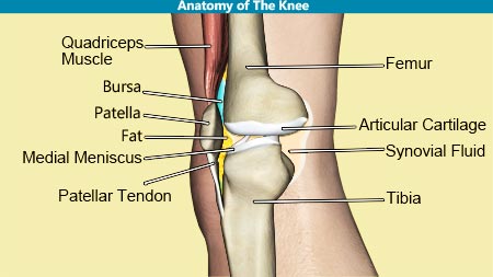
Knee joint is the largest joint in human body.1Body weight passes through knee joint during standing , walking , and ramble on . Knee spliff is used systematically during daily activities . Knee joint deteriorate following excessive use of human knee articulatio , harm , transmission , or kindling . Wear and split of stifle joint causes arthritis and ligamental sprain in later part of life . Knee marijuana cigarette is also known as hinge joint because of the movement like hinge of the door.2Knee joint links upper stage ( femur bone ) with the low leg ( tibia and fibula bone ) . The knee joint is form by femur , tibia , calf bone and kneepan ( knee cap ) . Knee joint controls movement of modest leg resulting in flexion and extension . Knee joint also achieves minimum external and inner rotary motion . Knee stick is a synovial spliff since the joint is enclosed within capsule of synovial membrane . The cartilages line the joint surface of the femoris , tibia , and kneecap . Knee joint is keep going by several tendon , ligament , musculus , and bursa .
Bones of Knee Joint
Four clappers take part in knee reefer : Femur , Tibia , Fibula , and Patella .
Femur-
Tibia-
Fibula-
Patella-
Knee Joint
Minor Knee Joint: Femoro-Patellar Articulation
The patella is also love as kneecap . The inner surface of kneepan is in contact with prior ( front ) open of lower end of femur cognize as patellar groove . Patella slide over patellar groove of femoris during flexure and extension . internal aerofoil of patella is line by a politic cartilage , which take into account smooth movement of kneepan over the anterior airfoil of femoris .
Major Knee Joint: Femoro-Tibial Articulation
thighbone is tie with tibia at median and lateral side . The median and sidelong articulating oddment of femur and tibia off-white are known as median and lateral condyle . sidelong condyle of femur is larger in sizing than median condyle . Articulating airfoil is cover with unruffled cartilages . sidelong condyle is spacious in front and medial condyle is unvarying in width from anterior to later surface . The smooth airplane and varying width of articulating control surface countenance roll movement to reach flection and extension of the knee stick as well as sliding movements .
Synovial Membrane
genu joint is also known as synovial articulation . Joint is covered from outside by a thin synovial membrane take shape a capsule .
Knee Joint Capsule
genu joint ejection seat is take form by synovial membrane and slender hempen layers .
Cartilages of Knee Joint
Articulating Cartilage-
Menisci-
median and sidelong menisci are made up of hempen gristle . open of meniscus are covered by hyaloid cartilage . Hyaline gristle provides smooth surface for slew and rolled movements of knee joint . Hyaline cartilages do not self - restore and are prone to wear and bout . Newly formed cartilages are mostly fibrous cartilage . Fibrous cartilages often build cracks and uneven surface do osteoarthritis .
Anatomy of Menisci-
Functions of Menisci-
Complications-
Cause of Complication-
Ligaments of Knee Joint
ligament are bundle of thick and tough fibrous tissue . Ligament links the bones to form a juncture .
Function of Ligament-
ligament of stifle joint are divided in to following 2 groups-
A. Intra - Capsular Ligament
B. Extra - Capsular Ligament
Anterior Cruciate Ligament (ACL)-
Posterior Cruciate Ligament (PCL)-
Transverse Ligament-
Anterior Menisco-Femoral Ligament-
Posterior Menisco-Femoral Ligament-
Menisco-Tibial Ligament (MCL)-
Lateral Collateral Ligament (LCL)-
Medial Collateral Ligament (MCL)
The MCL extends from the medial epicondyle of the femoris to the medial condyle and superior part of the median airfoil of the tibia . The MCL is steadfastly attach to the median meniscus , which is why these are commonly deplume at the same clip in contact sport .
Patellar Ligament
Ligament Retinacula
Oblique Popliteal Ligament
Arcuate Popliteal Ligament
Bursa of The Knee Joint
A. Anterior Group of Bursa
a. Suprapatellar Bursa
b. Patellar Bursa
B. Medial Bursa-
The Anserine Bursa
C. Posterior Bursa
The Semimembranosus Bursa
Tendons And Muscles of The Knee Joint
References :
Also Read :


